Morton's Neuroma Treatments
Morton's Neuroma Treatments
We always investigate the source and possible underlying causes for a neuroma. This may involve an in-depth assessment in our gait analysis facility or a specialist referral (to a neurologist, for example).
We are only happy to proceed with treatments such as cryosurgery
when we are satisfied that such investigations have taken place and that appropriate conservative care has been attempted.
Cryosurgery Treatment
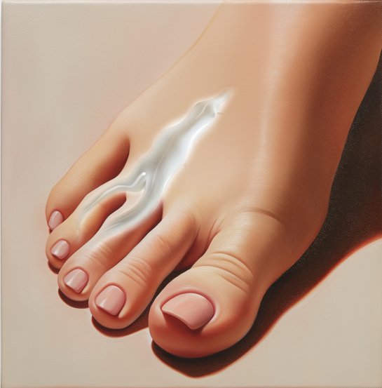
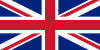
Cryosurgery involves freezing the neuroma by inserting a very thin needle-like probe. It is a precision procedure that requires a high level of technical skill and expertise in ultrasound. Cryosurgery is one of the most effective and safest available treatments, which is proven by our audited data. The London Podiatry Centre continues to lead the way in the management of Morton's neuroma. Most recently, Mr McCulloch has been a professional expert and advisor for the National Institute of Health and Clinical Excellence (NICE) in relation to the management of Morton’s neuroma. NICE is the key organisation that guides both NHS and private medicine and has acquired a high reputation internationally as a role model for the development of clinical guidelines. Mr McCulloch has acted as an expert both in relation to ablative radiofrequency, and most recently in June 2022, he was an expert in providing guidance on the use of cryosurgery for the management of Morton’s neuroma.
In cryosurgery, coldness is applied to abnormal tissue in order to resolve pain issues. Several different gasses such as nitrous oxide or CO2 can be used in this surgical procedure.
Cryosurgery has been used by pain specialists for many years to block the transmission of nerves in order to control pain. It is currently an effective treatment for such diverse conditions as skin abnormalities, cancers (prostate, liver, lung etc), Trigeminal neuralgia (chronic pain in the face) and cardiac arrhythmias (irregular heartbeat).
Whilst cryosurgery had been used to manage Morton's neuroma since the 1980's, high resolution ultrasound has allowed the treatment to become more effective.
The ‘Doppler’ function on an ultrasound scan allows the operator to see blood vessels that run next to the nerves, and therefore helps to locate the precise area for treatment.
Cryosurgery is highly reliant on the skill of the operator and requires expert control of both the ultrasound machine and cryoprobe (the tool used to perform cryosurgery). We do not advocate cryosurgery without ultrasound (known as ‘blind surgery’) as the results are less predictable.
The benefits of cryosurgery are clear. This minimally invasive procedure requires a much shorter period of post-operative rest and avoids the risks and much longer recovery of more invasive procedures.
We have audited our cryosurgery results extensively, and the procedure is successful in up to 80% patients.
To date (August 2025) no patient who has undergone cryosurgery at the Centre for Morton’s neuroma has experienced a serious or significant complication. In thousands of procedures, we have an extremely low infection rate.
Where the procedure has been successful, patients are generally happy to provide feedback by e-mail or phone and we may not need to see them again. Where patients still have some remaining discomfort, we will offer a further treatment, but only after three months have elapsed. If a patient fails to respond to cryosurgery, we generally offer radiofrequency before considering the option of open surgery.
Even though we have found complication rates to be very low, there are a number of possible risks with the procedure. The risks are as follows:
Infection:
In thousands of cases, we have encountered fewer than a handful of infections. These all settled rapidly with antibiotics. Our Centre has a low infection rate in general, and we have never seen serious hospital-based infections such as MRSA and C. Diff.
Thrombosis:
We have not encountered this at the Centre in the management of Morton's neuroma.
Recurrence:
Up to 80% of patients experience a successful outcome from a single cryosurgery treatment. Where the first treatment is partially effective, we will usually offer a second treatment, which often resolves the remaining symptoms.
Regression:
Cryosurgery is very unlikely to worsen your condition. In fact, we consider the risk of complication from steroid injection to be greater.
Complex pain syndrome:
This is a relatively rare condition (affecting just 0.02% of the population) characterised by an abnormal nerve related response to trauma, including surgery. The Centre has had one patient develop a mild form of this condition (approx 2001), but they made an excellent recovery.
Painful scar:
Because of the small incision (2-3mm) we have not encountered painful scarring and it would be highly unlikely to occur.
Haematoma formation:
This is a deep blister that can form following cryosurgery and radiofrequency. It is relatively common in cryosurgery and naturally breaks down with time. Given that the tissues are exposed to extreme cold, a degree of haematoma formation is quite normal.
Stump neuroma:
Stump neuromas can form when a nerve is cut or injured. It is a significant risk in open surgery, but we have not encountered this complication in cryosurgery. In fact, we have successfully treated patients with stump neuroma using cryosurgery.
NB: we attempt to regularly update these pages, but we ask all patients to double check with the Centre for the latest information on our complication rates. This information was last updated in August 2025.
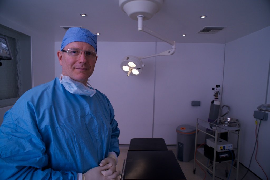
Ron McCulloch is a Consultant Podiatric Surgeon with over 30 years' of practical surgical experience. This means that he is qualified, certified and professionally endorsed to perform a full range of foot and ankle surgeries. Ron led the Podiatric Surgery department of Homerton NHS hospital for 10 years. During his time there, he trained fellow podiatric and orthopaedic surgeons in foot and ankle surgery, as well as lecturing extensively on foot and ankle pathology. When the Olympics came to London in 2012, Ron was called upon to offer his valued expertise to the world's athletes in the Olympic village. Other practitioners at the Centre have extensive experience and have undergone their advanced training with Mr McCulloch.
The London Podiatry Centre has been offering cryosurgery since 2011.
As demonstrated by our audit, the risks of complication is very low, especially for techniques such as cryosurgery. However, no surgery is entirely risk free. Patients can be reassured that Ron McCulloch has admitting rights to a number of hospitals, should medical admission be required after surgery.
The Morton’s Neuroma Centre is part of The London Podiatry Centre and is a CQC (Care Quality Commission) approved and regulated surgical clinic. The Care Quality Commission is the independent regulator of health and social care in England. Its role is to monitor, inspect and regulate services to make sure they meet fundamental standards of quality and safety. The commission publishes its findings, including performance ratings, to help people choose the best care. The CQC sets out what good and outstanding care looks like and makes sure services meet fundamental standards. You can read the CQC's latest audit report, published following their impromptu checks on provider services.
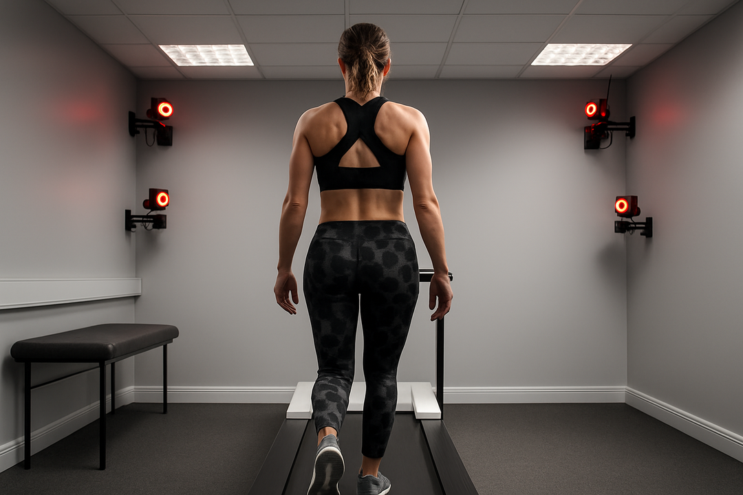
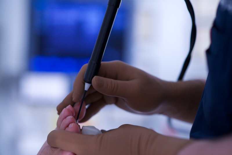
Certain foot conditions including Morton's neuroma are caused by a biomechanical weakness. These conditions have a moderate to high risk of recurrence when such biomechanical causes are not properly diagnosed and addressed. We have one of Europe's most sophisticated, dedicated podiatric gait analysis facility and have been trusted by Premiership football clubs and elite athletes since 1993. That means we can accurately diagnose and address the functional cause alongside treating the neuroma with cryosurgery. Treating the symptom and not the cause is often a false economy. Specialist practitioners extensively recognise that biomechanics must be treated when managing Morton's neuroma.
In the UK healthcare system, the title “Consultant” carries a very specific and significant meaning. A Consultant Podiatric Surgeon or Medical Consultant has held a substantive consultant post within the NHS, which means they have led a clinical service, supervised other clinicians, and taken full responsibility for patient care at the highest level. This title is not simply a job label, but a reflection of years of specialist training, formal accreditation, and senior leadership within a hospital setting. It’s important to distinguish this from the use of the term “consultant” in business or other industries, where it may be adopted more loosely without the same rigorous qualifications, clinical responsibility, or public accountability.
Ron McCulloch qualified as a Podiatric Surgeon in 2001 following principal roles at Guys and St Thomas', Lewisham and Homerton University Hospitals. He was appointed with the title Consultant Podiatric Surgeon in 2006. This means that Ron is a senior specialist with overall responsibility for patients’ care in our medical facility. Ron added the surgical techniques required for cryosurgery to his skill base in 2011 after studying under Professor Peter Wishnie in the United States. He has refined these surgical techniques ever since. Ron has a master’s degree in ultrasound and cryosurgery for Morton’s neuroma. He lectures and trains practitioners in the use of diagnostic ultrasound at Bournemouth University, Centre for Ultrasound Studies. Expertise in ultrasound is invaluable to correctly diagnose foot pathology and is crucial to performing cryosurgery for Morton's neuroma.
The London Podiatry Centre uses the Manchester Oxford Foot Questionnaire (MOXFQ) to audit the results of our surgical procedures including cryosurgery for Morton's neuroma. Validated for use in studies assessing outcomes following foot and/or ankle corrective surgery The MOXFQ allows us to provide clinically substantiated results for study and publication. We spend copious amounts of time obtaining clinically validated surgical data so our success rates are fully accurate and corroborated.
We provide definitive diagnosis and a full range of treatments for literally all foot and ankle ailments and injuries. You can therefore be assured that you will receive expert consultation and the relevant treatment regardless of your condition. In many cases, a previous misdiagnosis can lead patients to seek out cryosurgery. This is never a problem at the Centre as we can provide the full range of treatment for all foot-related issues, from conservative care through to full surgical management.
Mr McCulloch and the Centre are recognised by all major UK medical insurance providers and many international providers. Whilst requirements may vary among providers, medical insurers usually only accept the following criteria for professional recognition in Podiatric surgery:
• be a Fellow in Surgery of the Royal College of Podiatrists (FCR Pod (S))
• hold/have held a substantive post as a consultant podiatrist or consultant in podiatric surgery in the NHS
• be on the approved list of the Society of Chiropodists and Podiatrists
• have a practice that is not subject to any special conditions, restrictions or requirements for supervision or further training • be fully registered with the Health and Care Professions Council (HCPC)
• have current professional indemnity insurance for the treatments provided .
The efficacy and safety of cryosurgery for Morton’s neuroma are well-documented in peer-reviewed studies, which Mr McCulloch has summarised below:
Cazzato, R. L., et al. (2016).
Percutaneous MR-Guided Cryoablation of Morton’s Neuroma: Rationale and Technical Details After the First 20 Patients. Cardiovascular and Interventional Radiology.
Findings: In 20 patients (24 neuromas), MR-guided cryoablation achieved a 100% technical success rate, high patient satisfaction, and no major complications. The procedure took approximately 40.9 minutes on average, with minimal risk of stump neuroma. DOI: 10.1007/s00270-016-1365-7
Haddad, M., et al. (2024).
Safety and Efficacy of Percutaneous Morton Neuroma Cryoneurolysis Under Ultrasound Guidance. Cardiovascular and Interventional Radiology.
Page 2 of 2
Findings: A retrospective study of 54 patients (59 neuromas) reported a 98.1% technical success rate, no major complications, and only three minor complications (e.g., chilblain-type lesions). Six-month pain relief was promising, supporting cryosurgery’s safety and efficacy. DOI: 10.1007/s00270-024-03669-1
Friedman, T., et al. (2012).
Sonographically Guided Cryoneurolysis: Preliminary Experience and Clinical Outcomes. Journal of Ultrasound in Medicine.
Findings: In a case series including Morton’s neuromas, 15 of 20 patients responded positively to cryoneurolysis, with fewer adverse events compared to other ablation methods, highlighting its potential as a safe treatment option. DOI: 10.7863/jum.2012.31.12.2025
Caporusso, E. F., et al. (1997).
Cryogenic Neuroablation for the Treatment of Lower Extremity Neuromas. Journal of Foot and Ankle Surgery.
Findings: In 31 neuromas across 20 patients, cryosurgery achieved immediate pain relief in all cases, with 38.7% remaining pain-free at two weeks and 45.2% reporting reduced pain. Patient satisfaction was high (90%), with minimal disability. DOI: 10.1016/s1067-2516(97)80079-8
These studies collectively demonstrate cryosurgery’s superior outcomes compared to open surgery, which carries higher complication and dissatisfaction rates, often requiring costly follow-up investigations and pain management referrals.
Ablative Radiofrequency
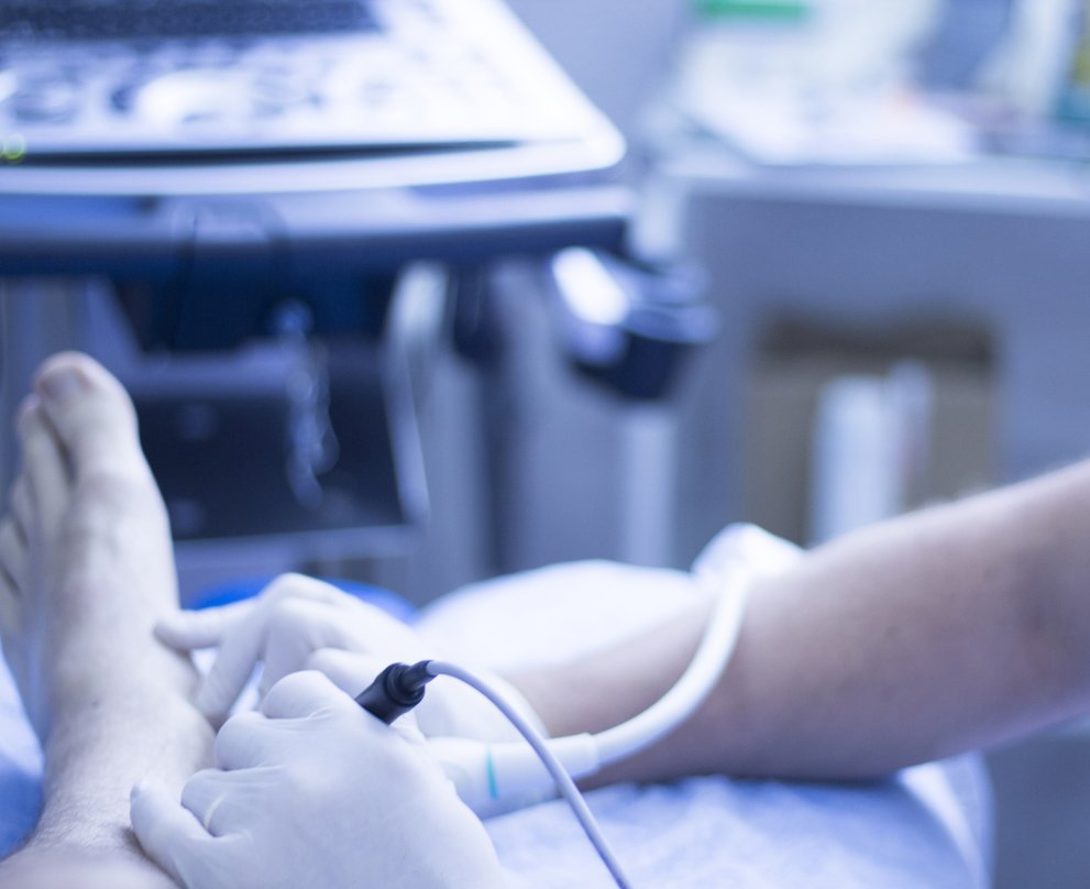
Ablative radiofrequency is similar to cryosurgery as it involves a needle being administered to the neuroma, guided by ultrasound.
However, rather than administering cold, heat is used to ablate (breakdown) the neuroma. Like cryosurgery, the procedure is performed under local anesthetic.
Radiofrequency causes a more permanent breakdown of the nerve. We generally recommend this procedure for patients who have already had cryosurgery but not experienced sufficient relief of symptoms.
a. The full procedure including anaesthesia usually takes no more than one hour.
b. We often use a tibial nerve block at the ankle to target the tibial nerve. Guided by an ultrasound machine to ensure a safe and accurate injection, this specialised local anaesthetic block brings a number of benefits including longer pain relief.
c. It generally takes no more than 20 minutes for the foot to go numb.
d. Using ultrasound guidance, we insert the radiofrequency probe and place it adjacent to the neuroma.
e. After starting the machine, we then administer a number of cycles.
f. A simple dressing is applied afterwards.
g. We advise patients to rest for 24 hours, after which they can start low-impact activities as long as they are comfortable doing so.
To date, none of our patients have experienced any significant complications from ablative radiofrequency treatment for Morton's neuroma.
Despite this, we believe that the procedure causes more tissue breakdown when compared to cryosurgery. Therefore, we generally recommend radiofrequency when cryosurgery and conservative care has not been successful.
Even though we have found complication rates to be very low, there are a number of possible risks associated with the procedure. These are explained further in the booklet we give to our patients before undergoing invasive treatment. These risks are as follows:
Infection:
None of our radiofrequency treatment patients have experienced this to date. Other studies have, however, recorded incidences of infection after the procedure.
Thrombosis:
We have not encountered this at the Centre. The risk of thrombosis is very low.
Recurrence:
When radiofrequency fails or recurs, options include a repeat treatment or open surgery.
Regression:
We have rarely seen patients worsen with radiofrequency, however this is possible.
Complex pain syndrome:
This is a relatively rare condition that is characterised by an abnormal nerve-related response to trauma, including surgery. We have not seen this condition in patients who have undergone radiofrequency.
Painful scar:
Because of the relatively small incision, none of our patients have experienced painful scars, and this would be highly unlikely to occur.
Haematoma formation:
This is a deep blister that can form after cryosurgery and radiofrequency. Haematomas are relatively common in cryosurgery and naturally break down with time. None of our patients have experienced haematomas after our radiofrequency treatment, but this complication could occur.
Permanent partial numbness of the one or more toes:
Whist our patients rarely encounter numbness with cryosurgery, this is more likely to occur to some extent with radiofrequency. Most patients tolerate this well and may not be aware that they have lost sensation.
NB: we attempt to regularly update these pages but please double check with the Centre for the latest information on our complication rates. This information was last updated in October 2018.
Radiofrequency can be repeated several times. Other forms of treatment should be considered if there is no discernible benefit after three treatments.
Recovery from radiofrequency treatment can take a little longer than cryosurgery. Whilst the procedure is sometimes associated with a little more discomfort, this is generally still very manageable. Most patients can return to work within 24 hours, especially if their work is sedentary. It takes about three months to determine whether the procedure has been successful.
Open Surgery
We will only offer open surgery to remove a neuroma in the few cases where conservative care, cryosurgery and radiofrequency have been unsuccessful.
We normally undertake a dorsal approach (top of the foot), and our success rate is over 90%. Procedures normally take place under local anaesthetic. Patients can generally return to work after two weeks, and sport after two months.
We prefer this treatment to some of the alternatives, including transverse ligament release and metatarsal osteotomy. The Morton's neuroma nerve only controls sensation, and not movement, and the majority of patients do not notice the slight loss of feeling that occurs after the operation.
This remains our preferred technique when conservative care and less invasive procedures like cryosurgery and radiofrequency have been unsuccessful. By making the incision on top of the foot, there is far less chance of a painful scar forming. We have relatively low complication rates with this procedure, although these are higher than cryosurgery and radiofrequency. The local anaesthetic is no more painful than a dental injection and is administered under ultrasound to ensure the best accuracy. The foot will generally stay numb for a minimum of six hours after surgery. However, most patients find that the local anaesthetic will last much longer. The procedure typically takes about half an hour and we remove the stitches at two weeks. After this time, patients can go back to work, however returning to sport activities is not advised for about two months. For patients who have previously had surgery, an incision at the bottom of the foot may be required to access the nerve. In these cases, the patient will not be able to weight bear for three weeks, which will help a smooth, fine scar to form that is unlikely to irritate. We use various specialised dressings to minimise the chance of painful scar formation. Our success rate from open surgery is high and none of our patients have encountered complications such as stump neuroma. However, there is a recognised increased risk with open surgery compared to less invasive treatments such as cryosurgery and radiofrequency. Recovery from open surgery also takes longer than these other treatments. For this reason, we would generally only recommend open surgery when cryosurgery and radiofrequency have been unsuccessful. Please bear in mind that at least 70 per cent of our patients have a successful outcome from cryosurgery and those who don't have a very good chance of responding to ablative radiofrequency. Therefore, very few of our patients require an open procedure.
This procedure involves removing the nerve (and often releasing the intermetatarsal ligament) through an incision underneath the foot. It is usually performed under local anaesthetic. Some surgeons prefer this procedure because the neuroma is closer to the underside of the foot and can therefore be located more easily. Also, a larger section of nerve can be removed as the nerve can be traced further back into the foot. Despite this, we will only perform a plantar incision during revision surgery if a dorsal surgical approach has previously failed. This is because a plantar incision can result in a painful scar that can often be more painful than the original neuroma. Such a scar can be very challenging to fix, although we use a number of specialist techniques to minimise the risk. This includes specific forms of plastic surgery suturing (stitching) and anti-scar dressings.
With this technique, a small incision allows us to release the intermetatarsal ligament. A Morton's neuroma forms because it is compressed against this ligament as the toes bend. We sometimes carry out a ligament release with the help of a small surgical camera. In some cases, we make a small incision by "feel" whilst in others we open the foot in order to see the ligament. Whilst positive results have been reported, we have some reservations about this procedure. Nerve decompressions are common in other parts of the body, but those parts are not compressed by a shoe and are therefore more likely to respond. The procedure involves cutting deeper tissue, which causes some scar formation. If unsuccessful, further surgery would increase scarring. In our opinion, performing a release in combination with excising the nerve maximises the chance of success. It is important to bear in mind that an open surgery procedure is only suitable for patients who have failed to respond to cryosurgery and radiofrequency, which suggests their problem is particularly resistant to treatment.
This operation involves cutting the metatarsal bone to reduce pressure on the neuroma. This type of surgery could be considered for patients who have a very narrow intermetatarsal space as confirmed by x-rays. However, we do not believe it should be used as a standard treatment for all forms of Morton's neuroma regardless of cause. A metatarsal osteotomy may be suited to patients who have suffered metatarsal bone fractures with secondary nerve compression. We tend to avoid performing lesser metatarsal osteotomies for Morton’s neuroma because they can be associated with an increased risk of transferring pain onto other metatarsals (transfer metatarsalgia) as well as other complications. This operation would not stop the transverse ligament from putting pressure on the nerve. It should therefore be considered in combination with a ligament release.
Alternative Treatments
We always investigate the source and possible underlying causes for a neuroma. This may involve an in-depth assessment in our gait analysis facility or a specialist referral (to a neurologist, for example).
We are only happy to proceed with treatments such as cryosurgery
when we are satisfied that such investigations have taken place and that appropriate conservative care has been attempted.
There is limited evidence to suggest that shockwave treatment works for Morton's neuroma. We offer shockwave therapy for a number of conditions, however not typically for Morton's neuroma as we remain unconvinced of its effectiveness.
See our causes page for the types of footwear that we recommend to help with Morton’s neuroma. We always recommend shoes based on each individual’s biomechanical requirement and whether the specific type of footwear is required for work, sport or general daily use. Our experts assess shoe function using the Centre’s state-of-the-art gait laboratory.
There is limited evidence to suggest that steroid injections have long-term benefits for Morton’s neuroma patients. However, some patients do find them helpful, especially if the condition is associated with a larger bursitis. We would only ever perform a steroid injection if there is no evidence of weakness of the joints that lie adjacent to the neuroma.
We usually recommend cryosurgery over steroid injections because it is associated with fewer risks and complications, and we have encountered many patients who have developed the more serious complication of a capsular (joint) tear following a steroid injection.
Steroid weakens the capsular tissue, especially if there is already an underlying degree of instability, for example from a hammertoe. Capsular tears can be more difficult to treat than neuromas and they nearly always result in a toe deformity.
Multiple steroid injections will also cause varying degrees of irreversible wastage and skin discolouration. Some practitioners may offer multiple steroid injections because they think this approach is the only viable option other than open surgery. This is simply not the case, and patients are much better off moving onto one of the other treatment options that we offer, which have less risk of complication.
Alcohol injections
Alcohol injections have provided benefit in some cases, evidenced by a limited number of studies. However, the injections are relatively painful and the alcohol cannot be kept local to the nerve, which increases the risk of adverse reactions to the adjacent tissue. We believe that alcohol injections should be avoided because of the risks of complication and our opinion is that the treatment simply isn't specific enough.
Phenol injections
Phenol injections are rarely performed for the condition as phenol will cause damage to adjacent tissues, including local blood vessels. We do not advocate this form of treatment because of the risk of complication. Studies have shown the technique to be effective in some cases (Magnan et al 2010).
Botulinum injections
Some early studies show that botulinum injections can have a positive effect on Morton’s neuroma symptoms. However, we do not currently recommend this approach due a lack of evidence. Botulinum has a temporary effect and patients are likely to require repeat injections.
Hydro-dissection
We use hydro-dissection in specific circumstances, usually after previous surgery. This relatively low-risk procedure involves using higher volumes of fluid (usually an anaesthetic) to treat scar tissue. It can be effective, as evidenced by the positive results we have had at the Centre. The accurate "flushing" of anaesthetic around the nerve can cause the release of adhesions and therefore reduce pain.
Whilst a limited number of studies suggest that intermetatarsal ligament release is successful in treating Morton’s neuroma, more evidence is required.
This treatment does not address the neuroma directly and in cases where the nerve is large, symptoms may continue. Unlike other parts of the body, where a nerve can be "surgically released", the foot is likely to experience some degree of continued compression because of footwear.
One risk of intermetatarsal release is that further surgery would be required if the procedure was unsuccessful, which would lead to increased scar formation within the foot.
Orthotic therapy, in combination with footwear, is one of the main conservative treatment options for managing Morton's neuroma. Bulky or large orthoses, or devices that do not accurately address the underlying biomechanical deficits, can actually make symptoms worse.
Computer-designed orthoses can be ultra-thin and optimised for accuracy, shoe fit and effectiveness. Orthoses work by stabilising the metatarsals and redistributing force, so that the nerve becomes less irritated. The Morton’s Neuroma Centre has one of the most advanced, dedicated podiatric gait laboratories anywhere in the world today. By scientifically analysing walking and running gait, it often becomes possible to manage the symptoms of Morton's neuroma. Gait analysis may lead to a wide variety of possible treatments and should be considered as a diagnostic test (just like an MRI scan or X-ray).